850140-73-7
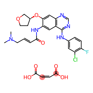
Afatinib Dimaleate
CAS: 850140-73-7
Molecular Formula: C28H29ClFN5O7
850140-73-7 - Names and Identifiers
850140-73-7 - Physico-chemical Properties
| Molecular Formula | C28H29ClFN5O7 |
| Molar Mass | 602.02 |
| Melting Point | >237oC (dec.) |
| Solubility | DMSO: ≥ 35 mg/mL |
| Appearance | Solid |
| Color | White to Pale Yellow |
| Storage Condition | Refrigerator |
| In vitro study | In lung cancer cell lines expressing wild-type (H1666) or L858R/T790M (NCI-H1975) EGFR, Afatinib was more effective in inhibiting cell growth than erlotinib, ge fi tinib and lapatinib. Afatinib was also effective on NSCLC cell lines expressing HER 2 776insV (NCI-H1781) or EGFR E746_A750del (HCC827), but not on A549 cells expressing wild-type EGFR and HER2. Afatinib enhanced the cytotoxicity of topotecan and mitoxantrone on SP cells and enhanced the apoptosis-inducing effect of topotecan and mitoxantrone on SP cells. |
| In vivo study | In a MDA-MB-453 xenograft model, Afatinib(20 mg/kg, P. O.) significantly induced tumor regression with a cumulative treated/control tumor volume ratio (T/C ratio). 2%, and down-regulation of EGFR and AKT phosphorylation levels. In A7, A431, FaDu, UT-SCC-14 and UT-SCC-15 xenograft models, Afatinib(30 mg/kg, oral administration) significantly prolonged tumor growth. In the HER2 amplified xenograft model, Afatinib(30 mg/kg, oral administration) significantly inhibited tumor growth and significantly prolonged survival. In the HER2 positive gastric cancer NCI-N87 xenograft model, oral administration of Afatinib(25 mg/kg) for 4 days significantly reduced the tumor volume and almost completely cured the tumor after 21 days of administration. |
850140-73-7 - Risk and Safety
| HS Code | 29339900 |
850140-73-7 - Preparation solution concentration reference
| 1mg | 5mg | 10mg | |
|---|---|---|---|
| 1 mM | 1.393 ml | 6.963 ml | 13.926 ml |
| 5 mM | 0.279 ml | 1.393 ml | 2.785 ml |
| 10 mM | 0.139 ml | 0.696 ml | 1.393 ml |
| 5 mM | 0.028 ml | 0.139 ml | 0.279 ml |
Last Update:2024-01-02 23:10:35
Supplier List
Spot supply
Product Name: Afatinib dimaleate Visit Supplier Webpage Request for quotationCAS: 850140-73-7
Tel:
Email: qianyanbiochem@gmail.com
Mobile: 13247110337
QQ: 2972965813
Product List: View Catalog
Spot supply
Product Name: BIBW2992 DiMaleate Visit Supplier Webpage Request for quotationCAS: 850140-73-7
Tel: +86-400-900-4166
Email: product@acmec-e.com
Mobile: +86-18621343501
QQ: 2881950922
Wechat: 19602116810
WhatsApp: +86-18621343501
Multiple SpecificationsSpot supply
Product Name: Afatinib Dimaleate Visit Supplier Webpage Request for quotationCAS: 850140-73-7
Tel: 0714-3999186
Email: 2853786052@qq.com
Mobile: 86+15671228036
QQ: 2853786052
Wechat: 15671228036
Product Name: BIBW2992 DiMaleate Request for quotation
CAS: 850140-73-7
Tel: +86 13454675544
Email: rachel@api-made.com
QQ: 3510434126
WhatsApp: +86 13454675544
CAS: 850140-73-7
Tel: +86 13454675544
Email: rachel@api-made.com
QQ: 3510434126
WhatsApp: +86 13454675544
Spot supply
Product Name: Afatinib (dimaleate) Visit Supplier Webpage Request for quotationCAS: 850140-73-7
Tel: 18301782025
Email: 3008007409@qq.com
Mobile: 18021002903
QQ: 3008007409
Wechat: 18301782025
Spot supply
Product Name: Afatinib dimaleate Visit Supplier Webpage Request for quotationCAS: 850140-73-7
Tel:
Email: qianyanbiochem@gmail.com
Mobile: 13247110337
QQ: 2972965813
Product List: View Catalog
Spot supply
Product Name: BIBW2992 DiMaleate Visit Supplier Webpage Request for quotationCAS: 850140-73-7
Tel: +86-400-900-4166
Email: product@acmec-e.com
Mobile: +86-18621343501
QQ: 2881950922
Wechat: 19602116810
WhatsApp: +86-18621343501
Multiple SpecificationsSpot supply
Product Name: Afatinib Dimaleate Visit Supplier Webpage Request for quotationCAS: 850140-73-7
Tel: 0714-3999186
Email: 2853786052@qq.com
Mobile: 86+15671228036
QQ: 2853786052
Wechat: 15671228036
Product Name: BIBW2992 DiMaleate Request for quotation
CAS: 850140-73-7
Tel: +86 13454675544
Email: rachel@api-made.com
QQ: 3510434126
WhatsApp: +86 13454675544
CAS: 850140-73-7
Tel: +86 13454675544
Email: rachel@api-made.com
QQ: 3510434126
WhatsApp: +86 13454675544
Spot supply
Product Name: Afatinib (dimaleate) Visit Supplier Webpage Request for quotationCAS: 850140-73-7
Tel: 18301782025
Email: 3008007409@qq.com
Mobile: 18021002903
QQ: 3008007409
Wechat: 18301782025
View History