287383-59-9
Scriptaid
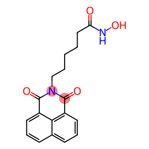
CAS: 287383-59-9
Molecular Formula: C18H18N2O4
287383-59-9 - Names and Identifiers
287383-59-9 - Physico-chemical Properties
| Molecular Formula | C18H18N2O4 |
| Molar Mass | 326.35 |
| Melting Point | 160-161 °C |
| Solubility | Soluble in DMSO (up to 4 mg/ml). |
| Appearance | Yellow solid. |
| Color | White |
| Storage Condition | -20°C |
| Stability | Stable for 2 years from date of purchase as supplied. Solutions in DMSO may be stored at -20° for up to 2 months. |
| Sensitive | Light Sensitive |
| Use | Scriptaid is a histone deacetylase (HDAC) inhibitor that can be used in cancer research. Scriptaid is also a sensitizer for antiviral drugs and can be used in the study of lymphoma associated with Epstein-Barr virus (EBV). |
| In vitro study | Scriptaid(6 μm) acted on PANC-1 cells to increase histone acetylation by more than 100-fold. Scriptaid(8 μm) is non-lethal to PANC-1 cells and has a limiting effect on MDA-MB-468 (80% survival). Scriptaid increases pCMVb, p6SBE-luc-and p6MBE-luc-independent transcription. Scriptaid can induce high expression of p6MBE-luc, pCMVb, and pUB6/V5-LacZ driven by viral (SV40 and CMV) or human (Ubiquitin c, UB6) promoters, this does not depend on the specificity of the enhancer (SBE and MBE), the type of promoter (viral and cellular), the product of the reporter gene (luciferase and β-galactosidase), and the integration status of the reporter gene structure. Scriptaid induces a high incidence of human nuclear transfer (SCNT) in oocytes developing to the blastocyst stage, and at all concentrations (50,100,250,500 and 2000 nm, respectively). All allowed their full-term development (3.4,4.2,7.6,6.8, and 4.1 percent, respectively). Scriptaid promoted the full-term development of cloned B6D2F1 embryos in a dose-dependent manner, with a maximum effect at 250 nm. Scriptaid clones all important inbred mouse strains such as C57BL/6, C3H/He, DBA/2, and 129/Sv. Scriptaid treatment of cloned embryos enhances the level of newly synthesized mRNA. Treatment of ICSI-fertilized embryos with 250 nM Scriptaid for up to 48 H did not inhibit development. Scriptaid inhibits the proliferation of T. gondii tachyzoite with an IC50 of 39 nM. Scriptaid(0.225 μm) completely protected HS68 monolayers from T. gondii tachyzoite invasion. Scriptaid inhibited the growth of ER-negative cell lines MDA-MB-231, MDA-MB-435, and Hs578t with an IC50 of 0.5-1.0 μg/mL after 48 h of treatment. 1 μg/ml Scriptaid treatment for 48 hours, induced the accumulation of acetylated H3 and H4 histone tail proteins, promoted ER mRNA transcription, and increased by 20000 times. Scriptaid inhibits the proliferation and viability of Ishikawa endometrial cancer cell line, and SK-OV-3 ovarian cancer cell lines with IC50 of 9 μm and 55 μm, respectively, while showing little sensitivity to normal endometrial epithelial cells. In the presence of Scriptaid, endometrial cancer cells and ovarian cancer cells were cultured for 2 days, and the cells were cultured in G0/G1 phase (5 μm Scriptaid treatment) and G2/M phase (10 μm Scriptaid treatment). Accumulation, while the S-phase proportion decreases accordingly. 10 μm Scriptaid induced apoptosis and mitochondrial membrane potential loss in 56.1% of Ishikawa cells, and decreased cyclin A and bcl-2 levels by 50% and 20%, respectively. |
| In vivo study | Treatment with Scriptaid at doses of 1.5 to 5.5 mg/kg elicited a dose-dependent reduction in lesion size (maximum 45% decline) and a subsequent reduction in motor and cognitive deficits. A similar protective effect can be achieved even if the treatment is delayed until 12 hours after injury. The protection of motor and cognitive function was long-lasting, with similar effects detectable at 35 days post-injury. Scriptaid treatment of the CA3 region of the hippocampus and pericontusional cortex induced an increase in viable neurons (42%), as well as an increase in their survival number/survival time. Scriptaid treatment of cortical and CA3 hippocampal neurons, inhibition of phosphorylated-AKT(p-AKT) and phosphorylated phosphatase, and TBI on chromosome 10 of the deleted tensin homolog (p-PTEN) decreased. A human breast cancer xenograft MDA-MB-231 model treated with Scriptaid at a dose of 3.5 mg/kg significantly inhibited tumor growth and reduced tumor volume by 75%. |
287383-59-9 - Risk and Safety
| WGK Germany | 3 |
287383-59-9 - Preparation solution concentration reference
| 1mg | 5mg | 10mg | |
|---|---|---|---|
| 1 mM | 3.064 ml | 15.321 ml | 30.642 ml |
| 5 mM | 0.613 ml | 3.064 ml | 6.128 ml |
| 10 mM | 0.306 ml | 1.532 ml | 3.064 ml |
| 5 mM | 0.061 ml | 0.306 ml | 0.613 ml |
Last Update:2024-01-02 23:10:35
287383-59-9 - Reference Information
| biological activity | Scriptaid is an HDAC inhibitor with a much higher effect on acetylated H4 than on H3. Scriptaid (GCK 1026) is an HDAC inhibitor with a much higher effect on acetylated H4 than on H3. |
| Target | Value |
Last Update:2024-04-10 22:29:15
Supplier List
Spot supply
Product Name: Scriptaid Visit Supplier Webpage Request for quotationCAS: 287383-59-9
Tel: +86-400-900-4166
Email: product@acmec-e.com
Mobile: +86-18621343501
QQ: 2881950922
Wechat: 19602116810
WhatsApp: +86-18621343501
Product Name: Scriptaid Request for quotation
CAS: 287383-59-9
Tel: +86 13313090628
Email: 13313091926@163.com
Mobile: +86 13313090628
Wechat: 13373390591
WhatsApp: 18733928930
CAS: 287383-59-9
Tel: +86 13313090628
Email: 13313091926@163.com
Mobile: +86 13313090628
Wechat: 13373390591
WhatsApp: 18733928930
Product Name: SCRIPTAID Request for quotation
CAS: 287383-59-9
Tel: +86 17733984678
Email: alice@jiuzhou-chem.com
QQ: 2284527519
WhatsApp: +86 17733984678
Linkedin: http://postmaster@api-made.com/
CAS: 287383-59-9
Tel: +86 17733984678
Email: alice@jiuzhou-chem.com
QQ: 2284527519
WhatsApp: +86 17733984678
Linkedin: http://postmaster@api-made.com/
Product Name: Scriptaid Visit Supplier Webpage Request for quotation
CAS: 287383-59-9
Tel: 18301782025
Email: 3008007409@qq.com
Mobile: 18021002903
QQ: 3008007409
Wechat: 18301782025
CAS: 287383-59-9
Tel: 18301782025
Email: 3008007409@qq.com
Mobile: 18021002903
QQ: 3008007409
Wechat: 18301782025
View History